Respiration
M. longicarpus
respires through the use of gills and complex brachiostegal lungs. This is
possible due to a modified gill chamber which is separated into two that allows
the circulation of water in one and air-breathing in another (Maitland &
Maitland, 1992). They are obligate air breathers as they obtain over 90% of
their oxygen requirement from their lungs (Maitland & Maitland, 1992).
Gills, sometimes referred to as branchia are a specialised
respiratory organ for large organisms that require oxygen extraction from water.
Crustaceans have internal gills that are composed of gill filaments which are
lined with high SA disk-like structures called lamella. Generally gills cannot
be used for air breathing, as they require the buyout medium of water to keep
gill filaments from drying out and collapsing onto one another (Hickman et al. 2006)
Unlike aquatic
crustaceans which mainly have nine pairs of gills, M. longicarpus has the
smallest number of gills recorded for any type of crab with only 5 gills
present(Farrelly & Greenaway, 1987). M.
longicarpus requires water to feed, so gill respiration is used during
feeding where water is absorbed by the setae of the abdomen from the substrate.
From here the water passes over the pericardial sacs, located side by side to
the carapace (Farrelly & Greenaway, 1987). These sacs act as an arrangement
of channels towards the capillary tubes (Mason, 1970). Once passed through the
perdicardial sacs the ventral margin of the epibranchial membranes forms the
“capillary tube” to which water passes over the gills. The lamella found on
gill filaments extracts oxygen from the water and uptakes it into its
circulatory fluid- hemolymph. Unique to the family Mictyris is the pattern of
haemolymph flow around the lamellae as all flow is firstly directed to the
marginal canal and is then directed into the lamellae central canal (Farrell
& Greenaway, 1987). The derived structure of M. longicarpus gill lamellae highlights the reduced importance of
gill respiration, with a reduction in the number of lamellae and the lamellae
spaced further apart (Farrell & Greenaway, 1987).
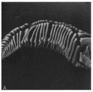
Picture of lamellae from the gill structure in M. longicarpus. Shows the few and far apart structure of the lamella. Image x15, taken from Farrelly & Greenaway (1987)
Branchiostegites,
which is an expanded branchial region of the carapace, divides the branchial
chamber into two, the inner branchial chamber is where the gills are located
and water is stored for feeding(Farrell & Greenaway, 1987). This leaves the outer branchial chamber to be
filled with air and function as a lung.
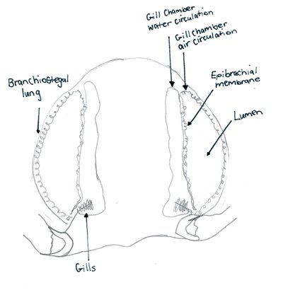
As adapted from Farrelly & Greenaway (1987), drawing of vertical section showing the composition of the two gill chambers in M. longicarpus. Drawing by Kate Buchanan, 2014.
The lung of M. longicarpus has an outer linning composed of epibranchial
membrane and has an inner linning of the branchiostegites (Farrelly &
Greenaway, 1987). As an increase in surface area increases the ability to
respire effectively, invagination of the lining appears within the lung to form
branching, blind ending pores, this makes the lung surface appear spongy
(Farrelly & Greenaway, 1987).
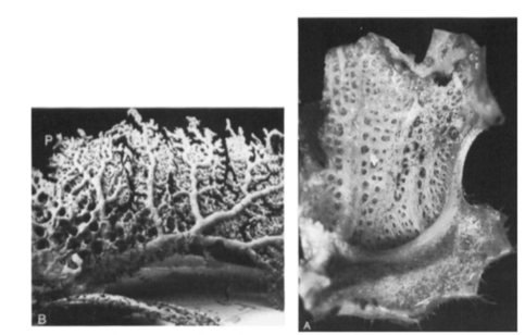
Left: a cast of the vasculature mainly of the epibranchial membrane viewed from the Lumen. Right: The spongy appearance of the removed branchiostegite viewed from the Lumen side. Both photographs taken from Farrelly & Greenaway (1987).
Air
flow into the lungs is supplied mainly via the eye sinuses. Three large vessels
are responsible for incoming air into the lung, these vessels further branch
off within the lung, and it is at the end of these blind branches where gas
exchange occurs (Farrelly & Greenaway, 1987).
|